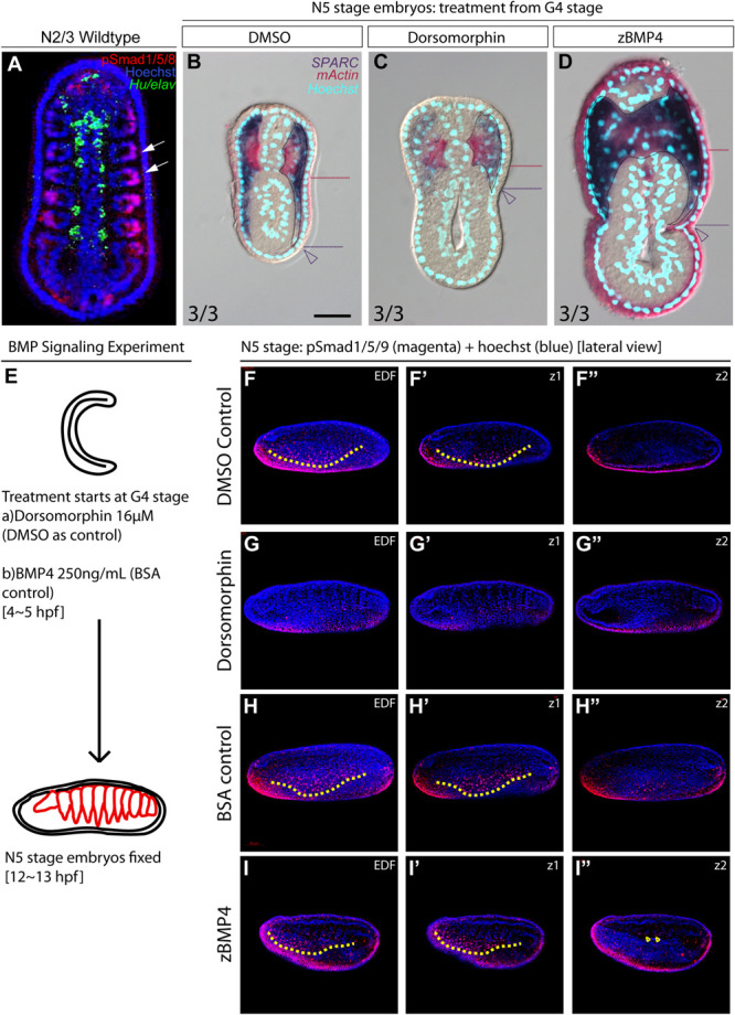
FIGURE 5.

Medial-lateral patterning of amphioxus somites is controlled by the level of BMP signaling. (A) Dorsal view of a stage N2/3 embryo. Anterior is to the top. Nuclear signals of pSmad1/5/8 are present preferentially in the lateral part of the somites (white arrows). Hu/elav (green) label indicates the midline structure in the embryo. (B–D) Cross sections of N5 embryos treated with BMP signaling inhibitor Dorsomorphin or recombinant zBMP4 protein at G4 stage. Scale bar, 25 μm. Red dotted lines mark the ventral boundary of the myotome region. Purple dotted lines mark the ventral boundary of the lateral somite region. Numbers on bottom left: denominator represents the total number of embryos that sectioned at 4∼6th somite level, numerator shows the number of embryos which show the displayed phenotype when sectioned. DMSO control is used as the count number for the control column. BSA whole mount embryos are shown in Supplementary Figure 4. (B) Double in situ hybridization showing the expression domain of SPARC and mActin in a control embryo treated with DMSO. (C) Section of N5 embryo treated with 16 μM dorsomorphin. (D) Section of N5 embryo treated with 250 ng/ml of zBMP4 protein. (B–D) Whole mount embryo of cross section in panels (B–D) is shown in Supplementary Figure 4. (E) Treatment time and concentration of BMP Signaling experiment. (F–I″) pSmad1/5/9 staining of embryos after BMP Signaling perturbation. Lateral view. Yellow dotted lines demarcate the ventral limit of the pSmad1/5/9 nuclear signal in the ventrally expanding somite. EDF, Extended Depth of Focus. Z1 demarcates the z-level which that corresponds to the EDF view. Z2 demarcates the z-level that is approximate at the notochordal level. (I″) Yellow arrowheads point to positive pSmad1/5/9 nuclear signal which roughly corresponds to the region where lateral gene markers are ectopically disorganized.
