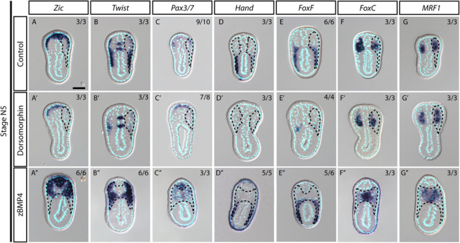
FIGURE 6.

Medial-lateral patterning of amphioxus somites is controlled by the level of BMP signaling. (A–G) Cross sections of N5 stage embryoes treated with DMSO as a control group. Scale Bar, 25 μm. (A′–G″) Cross sections of N4 stage embryos treated with inhibitor Dorsomorphin or recombinant zBMP4 protein at G4 stage. Dotted black outline marks one side of the somite. Whole somite areas are marked in panels (A″–G″), as left and right somites are not readily discernible. Left and right sides of somites exhibit left-right asymmetry as the left amphioxus somite approximately half a segment staggered more anteriorly to the right somite. Numbers on top right: denominator represents the total number of embryos that are sectioned at approximately 6th to 8th somite level as shown in Supplementary Figure 2, numerator shows the number of embryos which show the displayed phenotype when sectioned. DMSO control is used as the count number for the control column. BSA treated whole mount embryos are shown in Supplementary Figures 5–8. Whole mount embryos used from experiment are displayed in Supplementary Figures 5–8, sections are obtained from 6th to 8th pair somite, as indicated by the white brackets drawn in Supplementary Figure 2.
