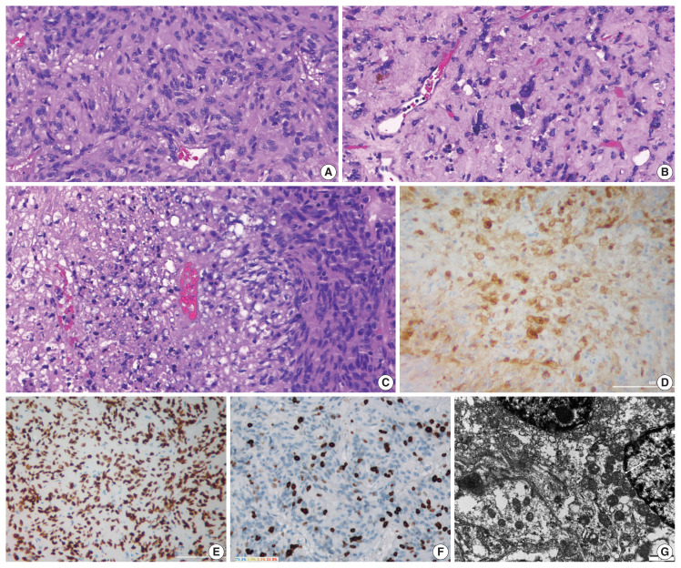
Fig. 3.
Histopathologic findings. (A) The tumor is composed of interlacing fascicles of spindle cells with abundant eosinophilic cytoplasm. (B) Focal nuclear pleomorphism and hyalinized stroma are seen. (C) Focal coagulative tumor necrosis is present. The tumor cells show immunoreactivity for galectin-3 (D) and thyroid transcription factor 1 (E). (F) The Ki-67 index was 26.7%. (G) Ultrastructural examination using paraffin-embedded tissue block show numerous mitochondria (white arrow) in tumor cell.

