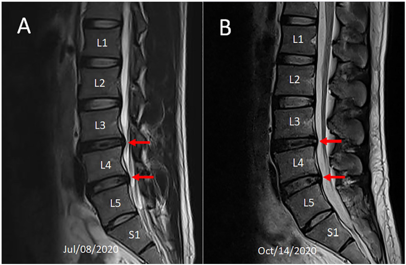
Figure 1.
Comparison of two MR scans over 3-month period. (A) Sagittal T2-weighted image before initiation of treatment showed decreased height of the L3/4, L4/5 and L5/S1 discs and reduced T2 weighted signal intensity (desiccation) of the L3/4 disc. Disc protrusion was seen at the L3/L4 and L4/L5 levels with indentation of the thecal sac (red arrows). (B) Follow-up image demonstrating regression of the thecal sac displacement (red arrows).

