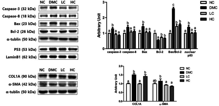
Figure 3.
Effect of vitamin D3 supplementation on hepatic apoptosis and fibrosis in type 2 diabetic mice. Representative images of Western blot for caspase-3, caspase-8, Bcl-2-associated X factor (Bax), Bcl-2, collagen 1 A (COL1A), and α-smooth muscle actin (α-SMA) in hepatic tissue from NC, DMC, and vitamin D3-treated mice (LC, 300 ng/kg of vitamin D3; HC, 600 ng/kg of vitamin D3). The band level of each marker was densitometrically quantified and normalized to that of α-tubulin (cytosol) and LaminB1 (nucleus). Data were presented as fold change relative to the NC group of mean ± S.E.M for each group (n = 6). Values with the different superscript letter were significantly different (P < 0.05; ANOVA with post hoc Duncan’s multiple range test).

