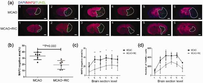
Figure 3.
Remote ischemic conditioning decreases infarction verified by MAP2 staining for live cells and neural cell apoptosis by TUNEL staining. (a) Representative images of brain tissue in the two groups as indicated (6 mice per group) with MAP2 and TUNEL immunofluorescence staining (scale bar, 1 mm) were depicted. The numbers of 1 to 6 represented coronal sections at different planes of each mouse brain. The brain tissue negative for MAP2 staining and positive for TUNEL (dead tissue) as marked by the white line was shown. (b) Average total infarct area (%) of six sections from each brain evaluated by MAP2 staining in the different groups was compared by the Student’s t-test. The infarct areas were calculated using the following formula: [(MAP2 positive area (live cells) of the nonischemic hemisphere) − (area of the MAP2 positive region in the ischemic hemisphere)/2 × area of the nonischemic hemisphere ×100%]. (c) Infarct area (%) evaluated by MAP2 staining in each of six sections was compared between the two groups by the Student’s t-test. (d) Apoptotic cells were evaluated by TUNEL immunofluorescent staining of different sections and counted for positive cells. The data were processed as described in the Materials and methods section and compared by the Student’s t-test. *p < 0.05, **p < 0.01. The similar findings were obtained when the same experiment was repeated independently.

