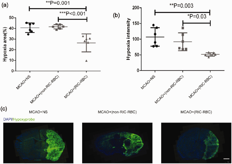
Figure 5.
Transfusion of 2,3-BPG-rich red blood cells prepared from RIC-treated mice increases the oxygen supply to ischemic brain tissue. (a) The average hypoxic areas (%) of all mice between the groups (6 mice per group) were compared by the one-way ANOVA followed by the post hoc test. The hypoxic area was calculated using the following formula: [area of nonischemic hemisphere (hypoxyprobe negative) − area of the hypoxyprobe negative region in the ischemic hemisphere]/2 × area of nonischemic hemisphere × 100%. (b) The average hypoxic intensities of all mice between the groups were compared by one-way ANOVA followed by the post hoc test. (c) Representative images of brain section at the same level (brain optic chiasm level) from each group were depicted (scale bar 1 mm). *p < 0.05, **p < 0.01, ***p < 0.001. The findings were similar when the same experiment was repeated independently. Twelve C57BL/6 mice were used as RBC donors for each experiment.

