Figure 3.
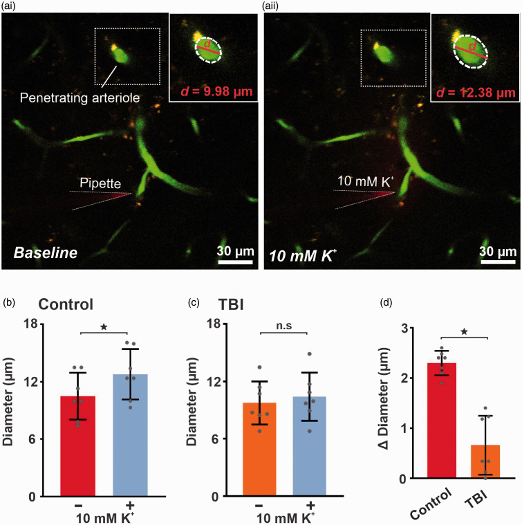
K+-induced capillary-to-arteriolar signaling is impaired in vivo after TBI. Micrograph illustrating pipette placement adjacent to a third-order capillary for in vivo monitoring of the diameter of the upstream feed arteriole (boxed) in (‘ai’) control and (‘aii’) TBI mice. Note the dilation in the penetrating arteriole (boxed). Magnification of the boxed areas around the penetrating arteriole, illustrate the magnitude of dilation evoked by capillary stimulation with 10 mM K+. Summary data showing arteriole diameter before and after capillary application of 10 mM K+, which produced significant upstream arteriole dilation when comparing diameters pre- and post-application of 10 mM K+ in (b) control (10 ± 2 vs 13 ± 3 µm, n = 7 paired experiments, 6 mice; *p < 0.0001, paired t test) but not in (c) TBI (10 ± 2 vs 10 ± 3 µm, n = 7 paired experiments, 6 mice; n.s., paired t test) mice. (d) The change in diameter after application of 10 mM K+ is significantly impaired in TBI (0.7 ± 0.6 µm, n = 7, *p < 0.001, unpaired t test) mice when compared to controls (2 ± 0.2 µm, n = 7). Data are expressed as mean ± SD.

