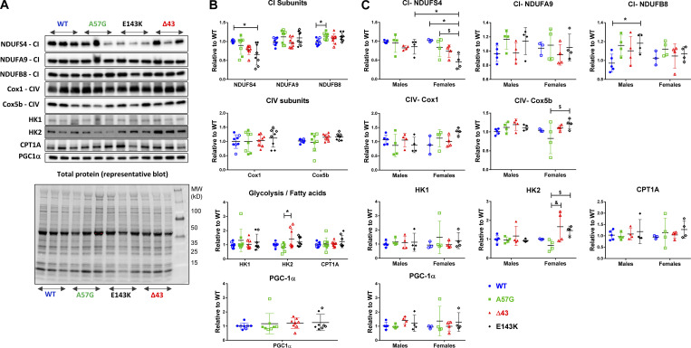
Figure S2.
Assessment of mitochondrial protein expression in heart homogenates from 7–11-mo-old HCM-A57G, Δ43, and RCM-E143K relative to WT-ELC mice. (A) Representative Western blots of mitochondrial and metabolism proteins in heart homogenates of mice. (B and C) Expression of CI, CIV, glycolysis, fatty acid subunits (HK1, HK2, and CPT1A), and peroxisome proliferator-activated receptor γ coactivator 1-α (PGC-1α; PPAR-γ coactivator and master regulator of mitochondrial biogenesis) for all four genotypes (B), and separately for female versus male mice (C). Values are mean ± SD of n = 8 (5 M, 3 F) WT-ELC; n = 8 (4 M, 4 F) HCM-A57G; n = 8 (4 M, 4 F) Δ43; and n = 8 (4 M, 4 F) RCM-E143K animals with significance depicted as *, P < 0.05 for mutant versus WT or between genders. $, P < 0.05 for HCM-A57G versus RCM-E143K; and &, P < 0.05 between HCM-A57G versus Δ43 analyzed by one-way ANOVA with Tukey’s multiple comparison test (B) and two-way ANOVA with two nominal variables (genotype, sex) followed by Tukey’s or Sidak’s multiple comparison test (C). Optical density was assessed with ImageJ. Each protein band was normalized to total protein content assessed for each blot before the primary antibody was applied. HK, hexokinase (glucose metabolism); NDUFS4, NDUFA9, and NDUFB8, CI subunits (NADH dehydrogenase); Cox5b and Cox1, CIV subunits; CPT1A, carnitine palmitoyltransferase 1A (fatty acid metabolism).

