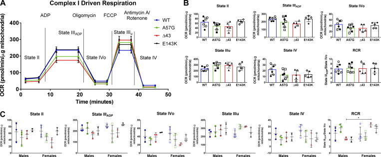
Figure S4.
CI-driven respiration profiles in the hearts of 8–10-mo-old WT, HCM-A57G, Δ43, and RCM-E143K mice. (A) Averaged traces of CI OCR in isolated mitochondria from WT-ELC (n = 7 mice), HCM-A57G (n = 7 mice), Δ43 (n = 5 mice), and RCM-E143K (n = 5 mice) using Seahorse XFp analyzer. (B) Characterization of mitochondrial OCR for state II, state IIIADP, state IVo, state IIIu, state IV, and the RCR, calculated as OCR-state IIIADP/OCR-state IVo. (C) Data from B separated by sex: WT (3 M, 4 F), HCM-A57G (3 M, 4 F), Δ43 (3 M, 2 F), and RCM-E143K (2 M, 3 F). Values are mean ± SD analyzed by two-way ANOVA with Tukey’s or Sidak’s multiple comparisons test with significance *, P < 0.05 for female versus male Δ43 mice. Open symbols depict female mice and closed symbols depict male mice.

