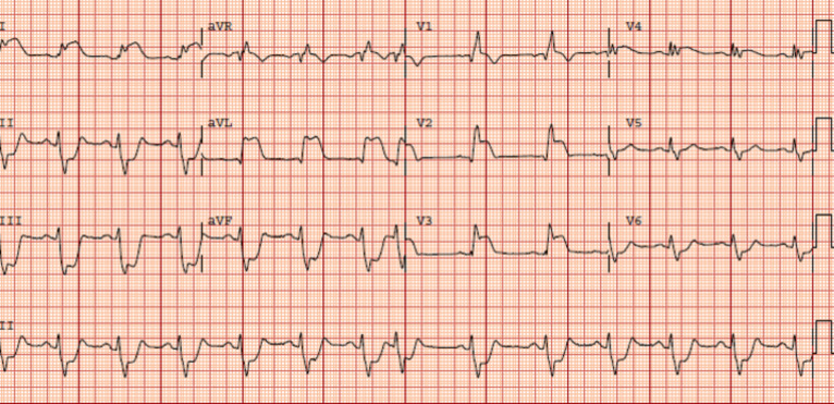
Fig. (3).
The ECG of a patient with proximal left anterior descending coronary artery occlusion: ST elevation in V1-V4, I and aVL and reciprocal ST depression in II, III, aVF, and V5-V6. There is a right bundle branch block and left anterior fascicular block. In addition, there is grade III ischemia (J-point /R wave ratio of >0.5 in I, aVL, V2-V3). (A higher resolution / colour version of this figure is available in the electronic copy of the article).

