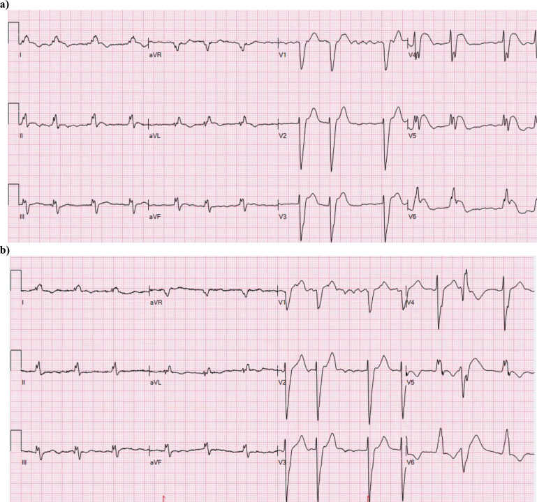
Fig. (5).
The ECG of a 74-year old man recorded 2.5 h after the initiation of chest pain. The medical history contains permanent atrial fibrillation with poorly controlled warfarin therapy, peripheral atherosclerosis, chronic obstructive pulmonary disease and frequent ventricular extrasystoles. The ECG in (a) shows LBBB with concordant ST elevation in I, and V5-V6. There is discordant ST elevation in V3-V4, but less than five mm. Coronary angiography showed single vessel disease with an occluded diagonal branch, possibly of embolic origin. (b) A previous ECG of the patient showing atrial fibrillation and LBBB (QRS 168 ms) with the “normal” secondary discordant ST/T changes associated with the conduction disorder. (A higher resolution / colour version of this figure is available in the electronic copy of the article).

