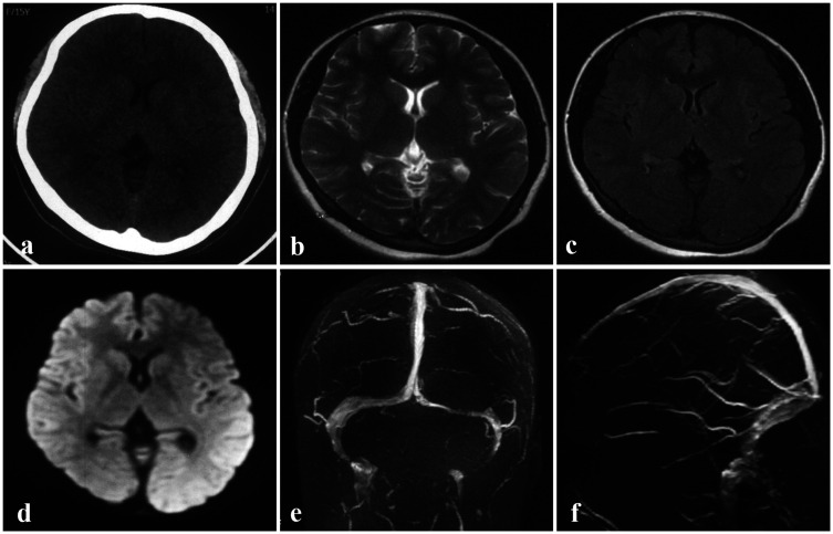
Figure 1.
Brain images obtained at a local hospital prior to admission. a–d: No cerebral parenchymal lesions were observed on NCCT (a), T2WI (b), FLAIR (c), or DWI (d). With the exception of the left slender TS, no other abnormalities were observed in the MRV maps (e–f).
DWI, diffusion-weighted imaging; FLAIR, fluid-attenuated inversion recovery; MRV, magnetic resonance venography; NCCT, non-contrast computed tomography; T2WI, T2-weighted imaging; TS, transverse sinus.

