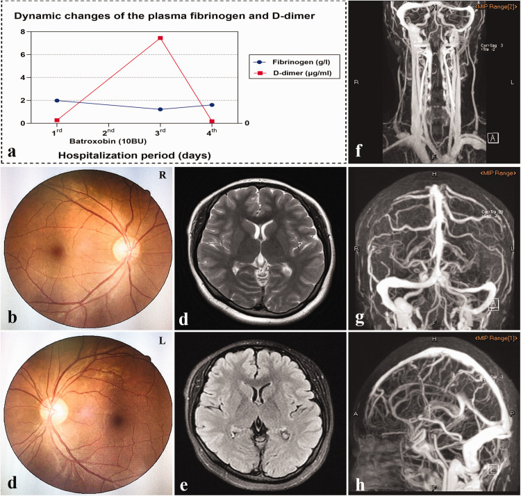
Figure 2.
Dynamic plasma fibrinogen and D-dimer, baseline fundoscopy examinations, and MRI and CE-MRV images during hospitalization. a: Dynamic plasma fibrinogen and D-dimer before and after batroxobin treatment. b, c: Bilateral fundus photographs revealed no papilledema (Frisen score = 0). d, e: No parenchymal lesions were observed in T2WI (d) or FLAIR (e). f–h: The left TS appeared slender and the venous vasculature of the entire brain was surrounded by dilated collaterals on the CE-MRV maps.
CE-MRV, contrast-enhanced magnetic resonance venography; FLAIR, fluid-attenuated inversion recovery; MRI, magnetic resonance imaging; T2WI, T2-weighted imaging; TS, transverse sinus.

