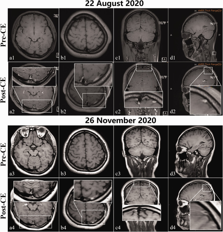
Figure 3.
Baseline (22 August 2020) and 3-month follow-up (26 November 2020) MRBTI maps. Hyperintensities within the left cortical veins, transverse/sigmoid sinus, and superior sagittal sinus on baseline CE-MRBTI sequences in axial (a2 and b2), coronal (c2), and sagittal (d2) sections were considered by a senior radiologist to be chronic venous thrombi. These hyperintensities had almost completely disappeared in the corresponding follow-up CE-MRBTI maps (a4–d4). The remaining hyperintensities in the left transverse/sigmoid sinus (b4) may be thrombosis-related venous wall inflammation. The shallow or disappeared cerebral sulci and fissures with unclear matter margins that were observed in the baseline CE-MRBTI (a1–d2) were deeper and more clearly observed in the follow-up CE-MRBTI (a3–d4), with well-defined matter borders.
CE-MRBTI, contrast-enhanced magnetic resonance black-blood thrombus imaging; MRBTI, magnetic resonance black-blood thrombus imaging; post-CE, post-contrast enhancement; pre-CE, pre-contrast enhancement.

