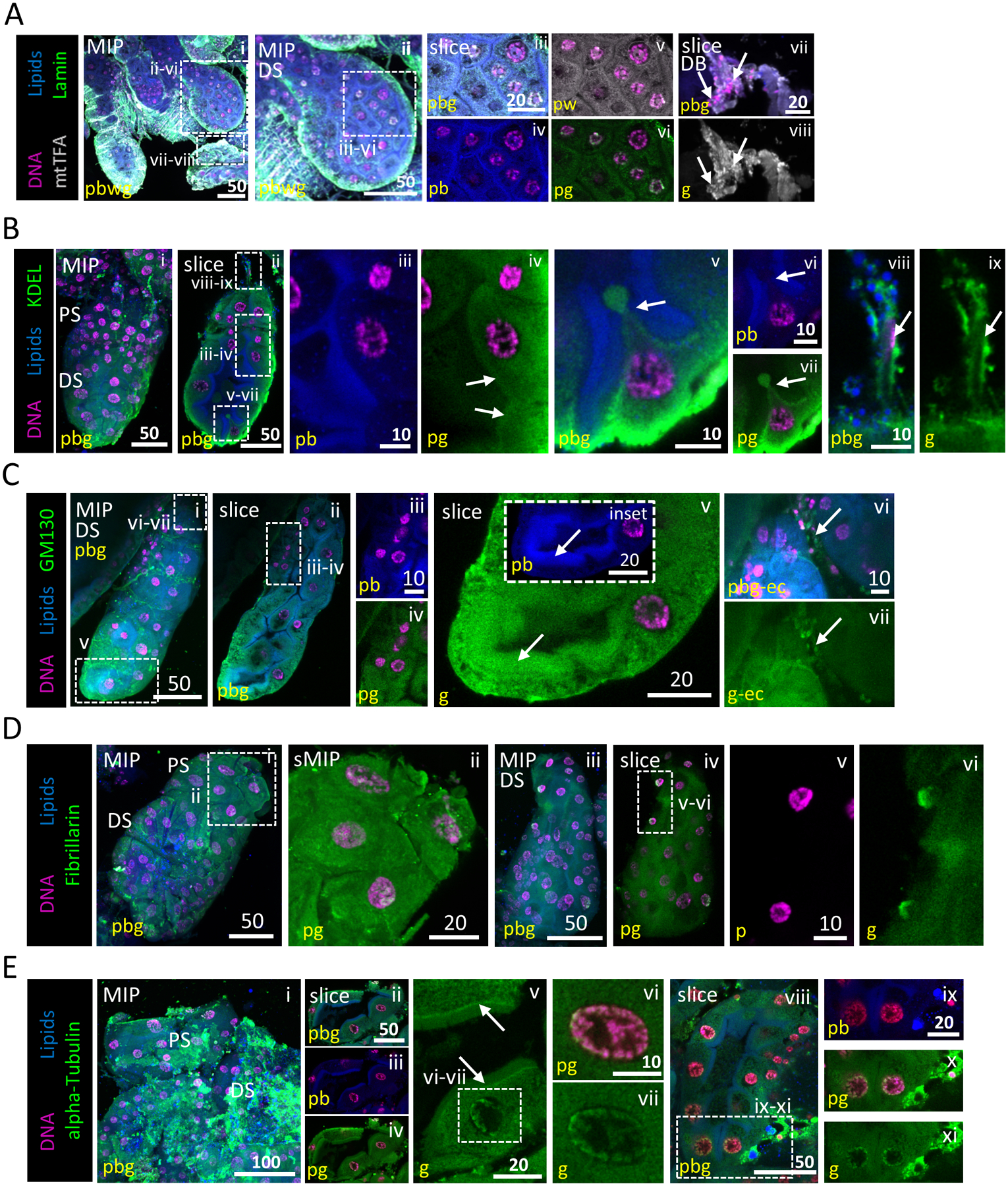
Figure 3. Subcellular organization of salivary gland cell organelles.

Representative images from dissected L4 larval salivary glands stained with Hoechst (DNA, purple), Nile Red (lipids, blue) and antisera against mtTFA (mitochondria, grey; A), lamin (nuclear membrane, green; A), KDEL (ER, green; B), GM130 (Golgi, grey; C), Fibrillarin (nucleolus, green; D) or alpha-tubulin (cytoskeleton, green; E). (A) mtTFA and lamin are broadly distributed within the cytoplasm. No intact nuclear envelope was observed in secretory sac nuclei (v). Some perinuclear lamin and mtTFA enrichment were observed, likely representing ER localization (iv, v). (B) KDEL is broadly distributed within the cytoplasm (ii, iv). A cell pinched between two adjacent apical brush border-like structures (v–vii) with a small cytoplasmic bud. Images from this L4 SG were also used in Fig. 5C. (C) GM130 is broadly distributed within cytoplasm (iv) and enriched (arrow, v) at the apical brush border-like structure (arrow, v inset). mtTFA (Avii, viii, white), KDEL (Bviii, ix, green) and GM130 (Cvi, vii, green) staining (arrows) are present in duct bud cells. (D) Fibrillarin staining is both diffuse cytoplasmic and in close association with multiple perinuclear sites (likely either fragmented nucleoli or ER association; ii, vi). (E) alpha-Tubulin staining is diffuse and striated through the secretory sac cell cytoplasm (i–iv). Signal enrichment was observed at the junction between the apical cytoplasmic face and the brush border-like structure (arrows, v). A halo of alphatubulin was seen in secretory sac nuclei, surrounding the chromosomes (v–vii, viii, x, xi).
