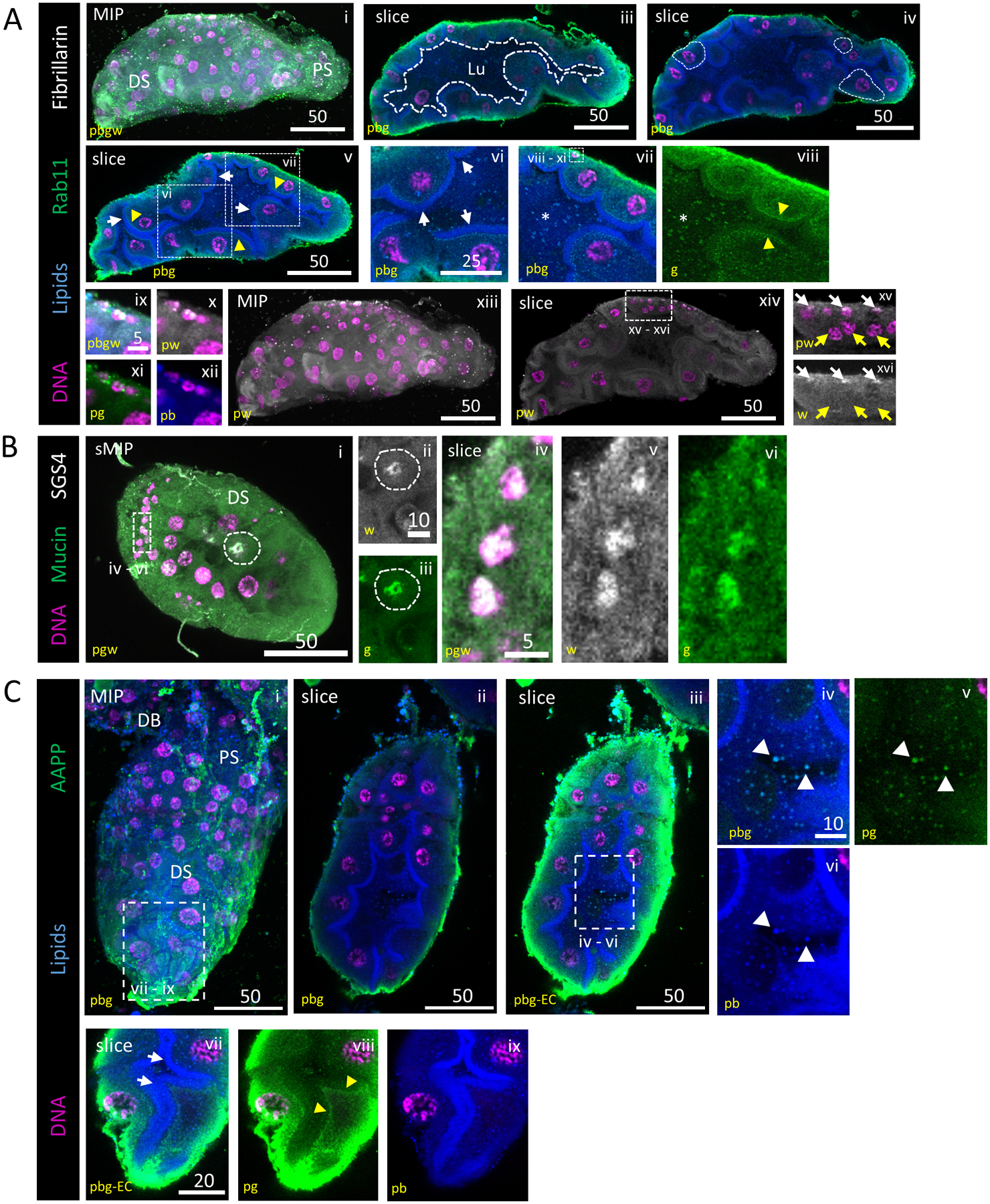
Figure 5. An apical brush border is visible during lumen formation and SG TFs localize to SG secretory and duct cell DNA.

Representative images from dissected L4 larval salivary glands stained with Hoechst (DNA, purple), Nile Red (lipids, blue) and antisera against Rab11 (vesicles, green) and either Fibrillarin (nucleoli, grey; A) or Sage (transcription factor, grey; B). (A) Secretory architecture of larval SGs. 3D and slice overviews are shown (i, ii). A growing, irregularly-shaped lumen (iii; white dashed outline) was seen. Larval SG cell size (iv; white dashed outlines) was variable. A brush border (vi; arrows), comprised of an apical lipid enrichment in many, but not all, cells, was present, confirming that these cells are polarized and specialized for secretion. Vesicle-like puncta were observed in the cytoplasm of secretory cells and in the lumen (vii; asterisk). Rab11-positive vesicles were enriched sub-apically, basal to the brush border (viii; arrowhead). Fibrillarin localization was only apparent in the smallest nuclei (ix–xvi) at the basal surface (xv–xvi; white basal arrows, yellow central arrows). (B) Mucin and SGS4(saliva proteins) staining were localized to a perinuclear enrichment, the broader secretory cell cytoplasm and the lumen. (C) AAPP, a saliva protein, is seen enriched at the basal surface of secretory cells, in intracellular vesicles and in extracellular vesicles (white arrows, iv–vi). AAPP-containing vesicles were seen enriched just basal to the apical lipid enrichment (yellow arrows, vii–ix). Different mages from this L4 SG were also included in Fig. S3B.
