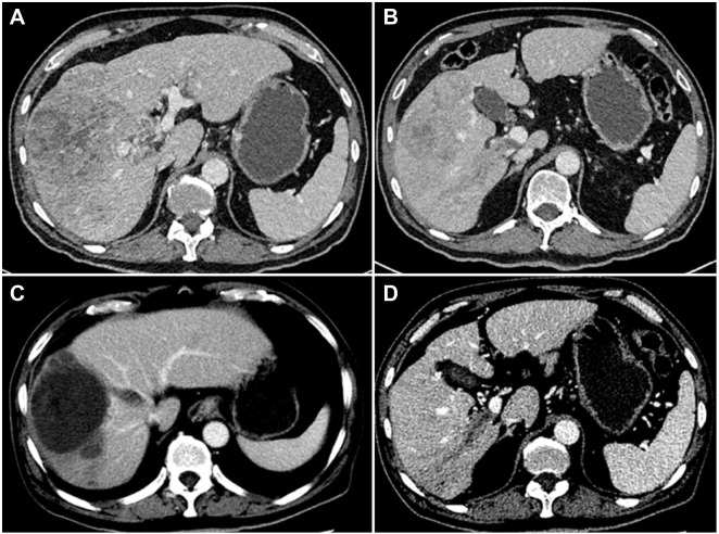
Figure 2.
Computed tomography image of the liver obtained from a 55-year-old male patient with a history of hepatitis B for 30 years. Contrast-enhanced CT imaging showed the presence of hepatocellular carcinoma with tumor thrombus in the right branch of the hepatic and portal veins (A and B). After 6 months of oral sorafenib combined with TACE, contrast-enhanced CT imaging showed tumor necrosis in the liver, and no blood supply was seen in the hepatic vein and PVTT (C and D).

