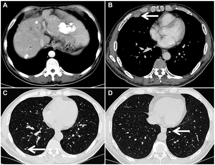
Figure 3.
Computed tomography images of the chest and liver obtained from a 51-year-old male patient who had been treated with transarterial chemoembolization alone for 7 months. Although the intrahepatic lesions were controlled (A), there were right-sided pleural metastases (B, arrow shown) and bilateral lung metastases (C and D, arrow shown).

