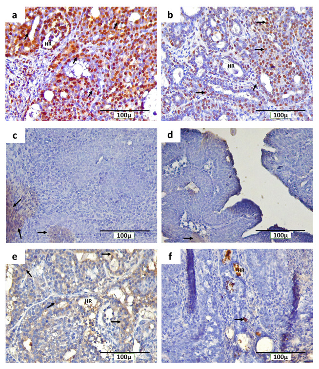
Figure 5. Immunohistochemical staining (3,3′-diaminobenzidine) of rat mammary tumors with PCNA, ErbB2, and caspase-3-positive cells.
( a) expression of PCNA in the nucleus of tumor cells in the non-therapy (INT) group, ( b) expression of PCNA in the nucleus of tumor cells in the therapy (IT) group, ( c) expression of ErbB2 on the cell membranes of tumor cells in the INT group, ( d) expression of ErbB2 on the cell membranes of tumor cells in the IT group, ( e) expression of caspase-3 in the cytoplasm of tumor cells in the INT group, ( f) expression of caspase-3 in the nucleus and hollow regions (HR) of tumor cells in the IT group. Brown color chromogen and arrows indicates positively stained cells.

