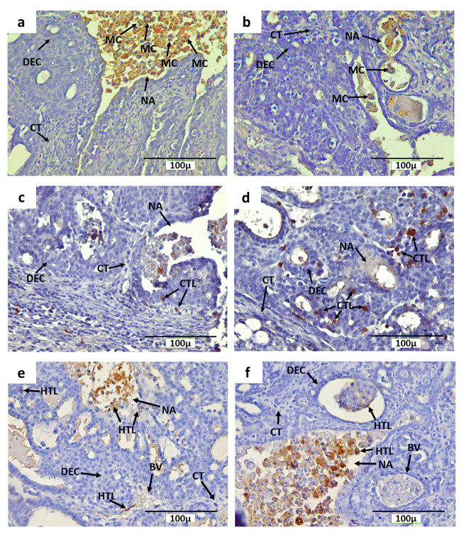
Figure 7. Immunohistochemical staining (DAB) of mammary tumors with CD68-positive macrophages, and CD4 and CD8-positive T cells.
( a) CD68 + macrophages in the non-therapy (INT) group, ( b) CD68 + macrophages in the therapy (IT) group, ( c) CD8 + T cells in the INT group, ( d) CD8 + T cells in the IT group, ( e) CD4 + T cells in the INT group, ( f) CD4 + T cells in the IT group. Here, brown color chromogen indicates positively stained cells. MC= macrophage cells, CT= connective tissue, BV= blood vessel, DEC= ductal epithelial cells, NA= necrotic area, CTL= cytotoxic T lymphocyte, and HTL= helper T lymphocyte.

