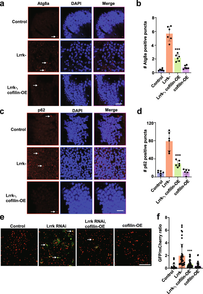
Fig. 7.
Autophagic pathology in Lrrk mutant flies is rescued by actin cytoskeletal destabilization. a, The number of Atg8a (LC3) immunoreactive puncta (arrows) is increased in the anterior medulla of homozygous Lrrk (Lrrke03680) mutant flies at 20 days of age. The increase in Lrrk mutants is reduced by expression of cofilin to destabilize the actin cytoskeleton, as quantified in (b). c, The number of p62-immunoreactive puncta (arrows) is increased in the medulla of homozygous Lrrk (Lrrke03680) mutant flies, and the increase in Lrrk mutants is reduced by expression of cofilin, as quantified in (d) when compared to control flies. Scale bars represent 10 µm in (a,c). n=6 per genotype. e, The number of GFP and mCherry puncta (arrows) is increased in flies with transgenic RNAi knockdown of Lrrk also expressing the GFP-mCherry-Atg8a reporter, and decreased by expression of cofilin. Scale bar represents 10 µm in (e). f, Quantification of the ratio of GFP to mCherry fluorescence indicates decreased autophagic flux in brains of flies with Lrrk knockdown mediated by transgenic RNAi, and partial normalization by overexpression of cofilin when compared to flies expressing transgenic Lrrk RNAi. n=4 per genotype. Data are represented as mean ± SD. ***p<0.005, ANOVA with Bonferroni post-test analysis. Control is nSyb-GAL4/+ in (a-d) and UAS-GFP-mCherry-Atg8a/+; nSyb-GAL4/ + in (e,f). Flies are 20 days old

