
Figure 2. Astrocytes interact with remodeling vessels after stroke.
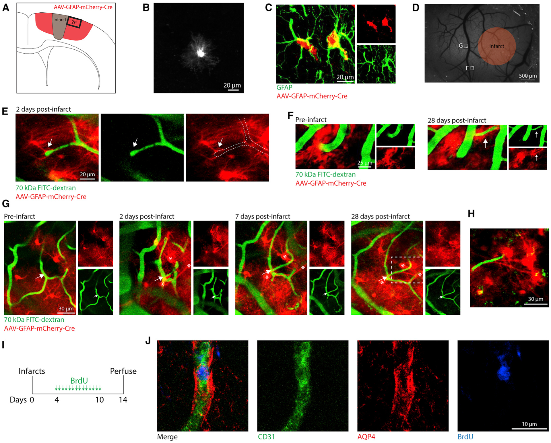
(A) Illustration of AAV-mediated labeling and imaging.
(B) In vivo two-photon (2P) image of an AAV-labeled cell with characteristic astrocyte morphology (pre-stroke).
(C) Confocal image validating AAV-mediated labeling of astrocytes with immunohistochemistry.
(D) Wide-field speckle contrast image of the brain surface. Orange region represents the approximate boundaries of the infarct core. White boxes indicate 2P-imaged regions corresponding to labeled images.
(E) 2P image from 2 days post-stroke of a vascular sprout contacted by a nearby astrocyte (arrow). This region was only imaged at 2 days post-stroke.
(F) 2P images showing a vessel that reoriented and increased diameter after stroke ensheathed by astrocytes (arrow).
(G) 2P images showing a new capillary segment (arrow) formed after stroke contacted by astrocytes (*).
(H) 2P image corresponding to the dashed box in (G) showing astrocytes extending processes that terminate on the new vessel segment.
(I) Experimental design for labeling proliferating vessels after stroke.
(J) Confocal images showing a new vessel (CD31+BrdU+) ensheathed by astrocytic endfeet (AQP4+). All CD31+BrdU+ new vessels analyzed (76/76 new vessels from six mice) were surrounded by AQP4+ endfeet.