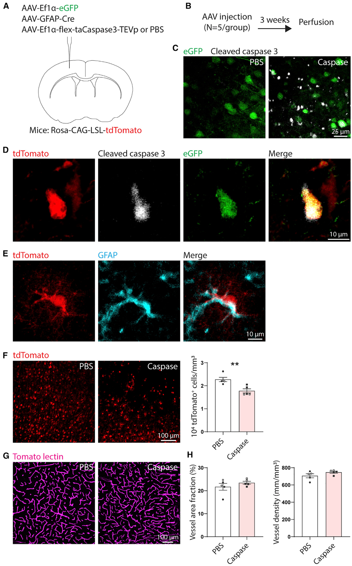
Figure 6. Genetic ablation of astrocytes in the otherwise intact brain does not affect vascular structure.

(A) AAV injection diagram. n = 5 Rosa-CAG-LSL-tdTomato mice per group (PBS or caspase). Mice in both groups were intracranially injected with AAV-EF1α-EGFP to label the injection site and AAV-GFAP-Cre to express Cre in astrocytes. Mice in the caspase group also received AAV-EF1α-flex-taCaspase3-TEVp to induce Cre-dependent expression of cleaved caspase-3, which causes apoptotic cell death. Control mice were injected with an equal volume of PBS instead of AAV-EF1α-flex-taCaspase3-TEVp.
(B) Experimental timeline.
(C) Confocal images showing expression of cleaved caspase-3 labeled by immunohistochemistry in mice injected with AAV-EF1α-flex-taCaspase3-TEVp (caspase), but not PBS. Images were taken at the injection site (EGFP label).
(D) Example of a tdTomato+ cleaved caspase-3+EGFP+ cell (caspase group). Images are 5-μm maximum projections.
(E) Example of a tdTomato+ GFAP+ cell, confirming Cre-mediated recombination in astrocytes.
(F) The number of tdTomato+ cells was reduced in the caspase group, confirming ablation. Images are 6-μm maximum projections. **t(8) = 3.82, p = 0.005, t test.
(G) Representative maximum projection confocal images of tomato lectin-labeled vasculature at the injection site from caspase AAV- and PBS-injected animals.
(H) Ablation of astrocytes did not affect vascular area fraction or density (t(8) ≤ 1.6, p ≥ 0.146, t tests). Data are presented as mean ± SEM. Data points representing males are shown as filled symbols; data points representing females are shown as open symbols.
