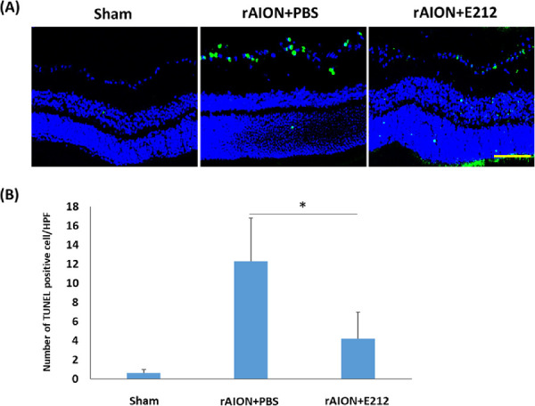
Figure 5.

Analysis of RGC apoptosis in the RGC layer through the TUNEL assay at week 4 after rAION induction. (A) Representative images of apoptotic cells in the RGC layers in the sham, PBS-treated, and E-212-treated group at week 4 after AION induction. The TUNEL-positive cells (green) and the nuclei of RGCs (blue) were stained with DAPI staining. (B) Quantification of TUNEL-positive cells per HPF. The number of apoptotic cells in the E-212-treated group were significantly lower (2.93-fold, *P < 0.05, n = 6 per group, scale bar: 100 µm) than that in the PBS-treated group. Data are expressed as the mean ± SD. HPF, high-power field; rAION, rodent model of anterior ischemic optic neuropathy; RGC, retinal ganglion cell; TUNEL, TdT-dUTP nick end-labeling.
