Figure 1.
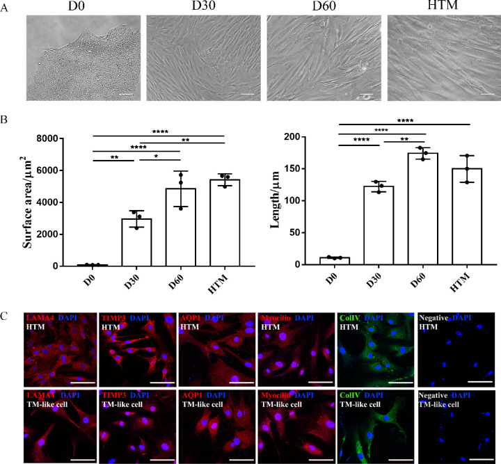
Characterization of human TM-like cells induced by the conditioned medium. (A) Morphological observations of TM-like cells during differentiation. Human iPSCs colonies of U1 were generated via reprogramming renal urethra epithelial cells. Through co-culturing with HTM cells, iPSCs were differentiated for 60 days. The morphological changes monitored by Nikon microscopy were compared with HTM cells of donor 1. (B) Surface area and length of TM progenitor and TM-like cell. TM progenitors (D30) were significantly smaller than TM-like cells (D60). Tukey's multiple comparisons test was used for data analysis. *P < 0.05, **P < 0.01, ***P < 0.001, ****P < 0.0001. (C) Immunohistochemical characterization of TM-like cells differentiated for 60 days. Laminin A4 (LAMA4), tissue inhibitor of matrix proteases 3 (TIMP3), aquaporin 1 (AQP1), myocilin (MYOC), and collagen type IV (ColIV) were detected with robust expression in both TM-like cells and HTM cells. Scale bar: 100 µm.

