FIGURE 2.

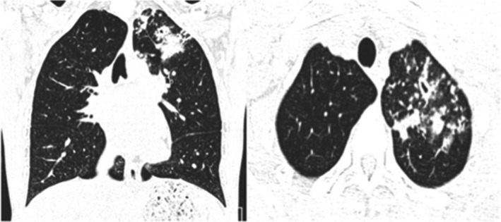
Atypical CT imaging features for COVID‐19. Axial (A) and coronal (B) CT images showing unifocal patchy consolidation with tree‐in‐bud opacities and centrilobular nodules

Atypical CT imaging features for COVID‐19. Axial (A) and coronal (B) CT images showing unifocal patchy consolidation with tree‐in‐bud opacities and centrilobular nodules