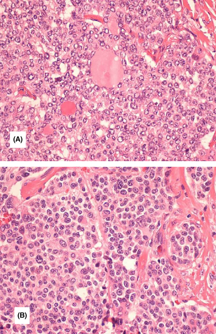
FIGURE 1.

Thyroid section showing mixed medullary and papillary thyroid carcinoma. A, Cells with characteristics of papillary thyroid carcinoma (irregular nuclear membrane, nuclear overlap and clarified chromatin), sometimes with follicle formation. B, Interspersed with salt and pepper chromatin cells. Histological type (WHO): Mixed carcinoma (composed) by a component of medullary carcinoma and another of follicular cells with characteristics of papillary carcinoma in the left lobe; In the right lobe, a 1 mm papillary microcarcinoma is observed. Multifocality/intrathyroid spread: observed, with sizes from 1 to 23 mm. Vascular and lymphatic invasion: Observed. Infiltrative pattern. Invasion of the gland capsule: Observed Extra‐thyroid extension: Observed; Limited, however, without involvement of striated muscle tissue. Margins: Not involved by neoplasia. Lymph nodes: Metastases (from the medullary and papillary components) are observed in 12 of the 38 lymph nodes isolated on the left and 8 of the 10 in the central region; Size of the largest metastasis: 20 mm—T2N1BM0
