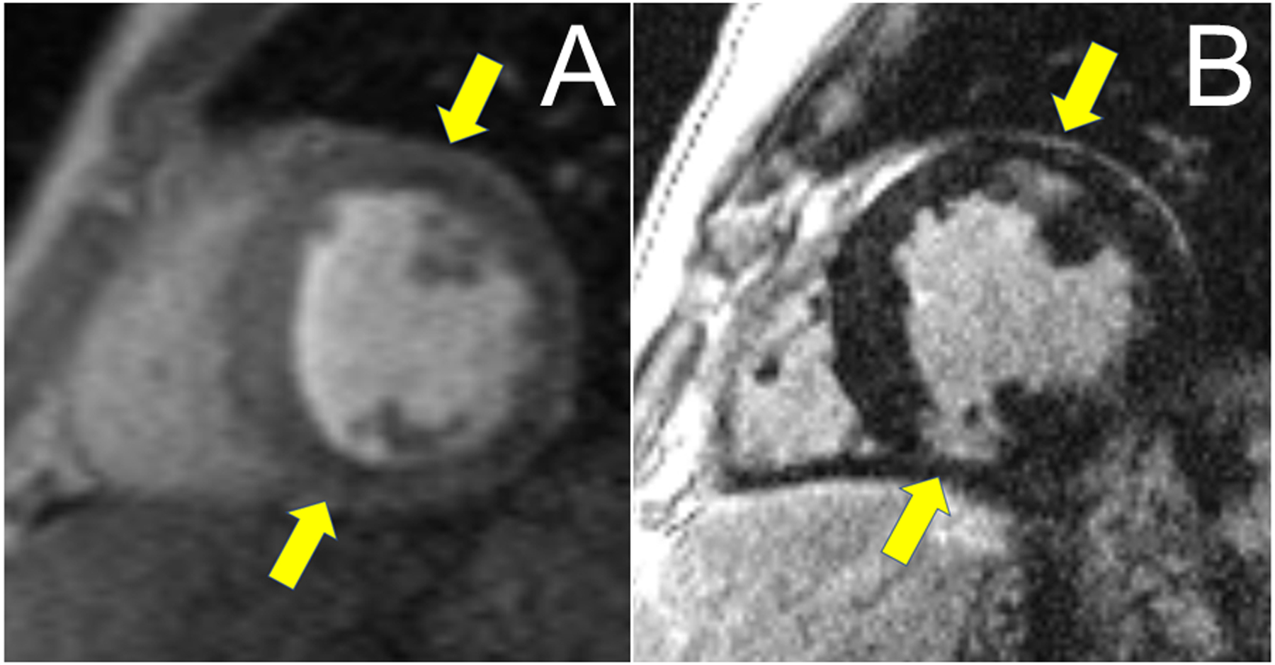
Figure 3:

Panel A: CMR with areas of subtle hypoperfusion seen in Gradient Echo Perfusion Imaging (GRE) as areas of decreased signal intensity in endo and mid-myocardium of the LV (yellow arrows). Panel B: Areas of corresponding LGE seen in the anterior (endo/midmyocardial) and inferior (near transmural) wall of the LV (yellow arrows).
