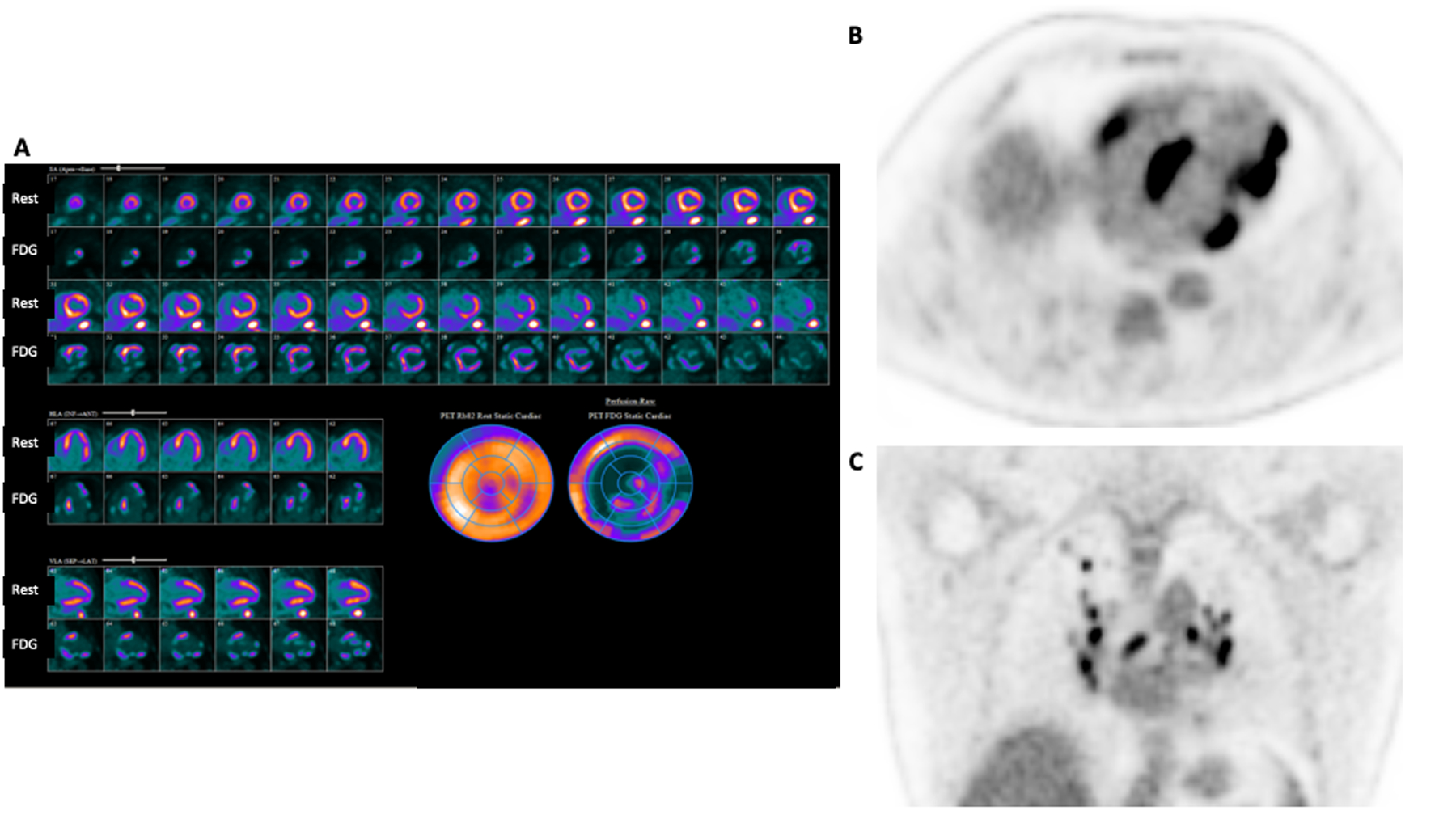
Figure 4:

Rest 82-Rubidium perfusion images from Case 2 show perfusion defects in the basal septum, basal anterior and anterolateral segments, as well as apical inferior, apical lateral, and mid inferolateral segments suggesting areas of scar (A). 18F-FDG PET imaging shows a mismatch pattern with increased uptake in these segments, increased uptake in adjacent basal and mid inferior as well as mid anterolateral segments (A), and increased RV uptake (B) consistent with active inflammation in these areas. The combination of perfusion defects (scar) and 18F-FDG uptake (inflammation) allows a more complete understanding of the patient’s burden of disease, prognosis, and pathology. Whole body imaging (C), shows increased 18F-FDG uptake in hilar and mediastinal lymph nodes.
