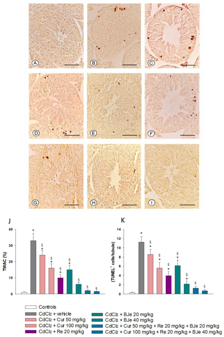
Figure 4.
Assessment of apoptosis in mice testes with TUNEL staining technique (scale bar: 50 µm). (A): In control mice, no TUNEL-positive cells can be observed. (B): In the seminiferous epithelium of mice challenged with CdCl2 alone, a large number of peripheral TUNEL-positive germ cells are present. (C): Mice challenged with CdCl2 and treated with Cur at 50 mg/kg. Many TUNEL-positive germ cells are observed. (D): Mice challenged with CdCl2 and treated with Cur at 100 mg/kg. A reduction of TUNEL-positive cells is evident. (E): Mice challenged with CdCl2 and treated with Re. A further reduction in the number of TUNEL-positive cells is evident. (F): CdCl2 plus BJe in 20 mg/kg-treated mice. TUNEL-positive cells are more numerous. (G): In CdCl2 plus BJe alone in 40 mg/kg-treated mice, the number of TUNEL positive cells is significantly reduced. (H,I): In both extract associations Cur 50 mg/kg + Re 20 mg/kg + BJe 20 mg/kg and Cur 100 mg/kg + Re 20 mg/kg + BJe 40 mg/kg-treated mice, isolated or even no TUNEL-positive germ cells are observed in the periphery of the seminiferous tubules. The images are representative of the nine mice of each experimental group. (J): Tubules with apoptotic cells (TWAC) (expressed in %) in the different groups of mice. (K): Apoptotic index (apoptotic cells/tubule) in the different groups of mice. * p < 0.05 versus controls; § p < 0.05 versus CdCl2-treated mice.

