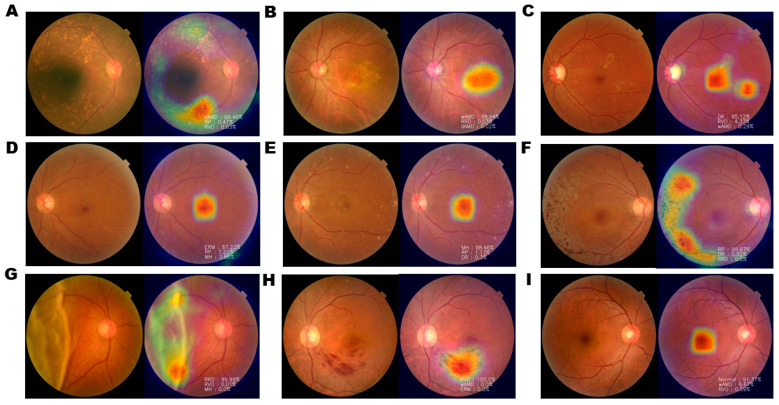
Figure 2.
Examples of Gradient-weighted Class Activation Mapping (Grad-CAM) visualization of retinal diseases. Grad-CAM visualization of (A) dAMD, (B) wAMD, (C) DR, (D) ERM, (E) MH, (F) RP, (G) RRD, (H) RVO, and (I) normal retina. Grad-CAM extracts the feature map of the last convolution layer and shows a heatmap within the image describing the calculated weight of the feature map. Heatmap images of nAMD show that the AI tool identified pathological changes, such as drusen, bleeding, elevation of the center, pigmentation, surface wrinkling, and retinal detachment. However, in normal controls, the center of macula is identified, with no degenerated area.

