Figure 2.
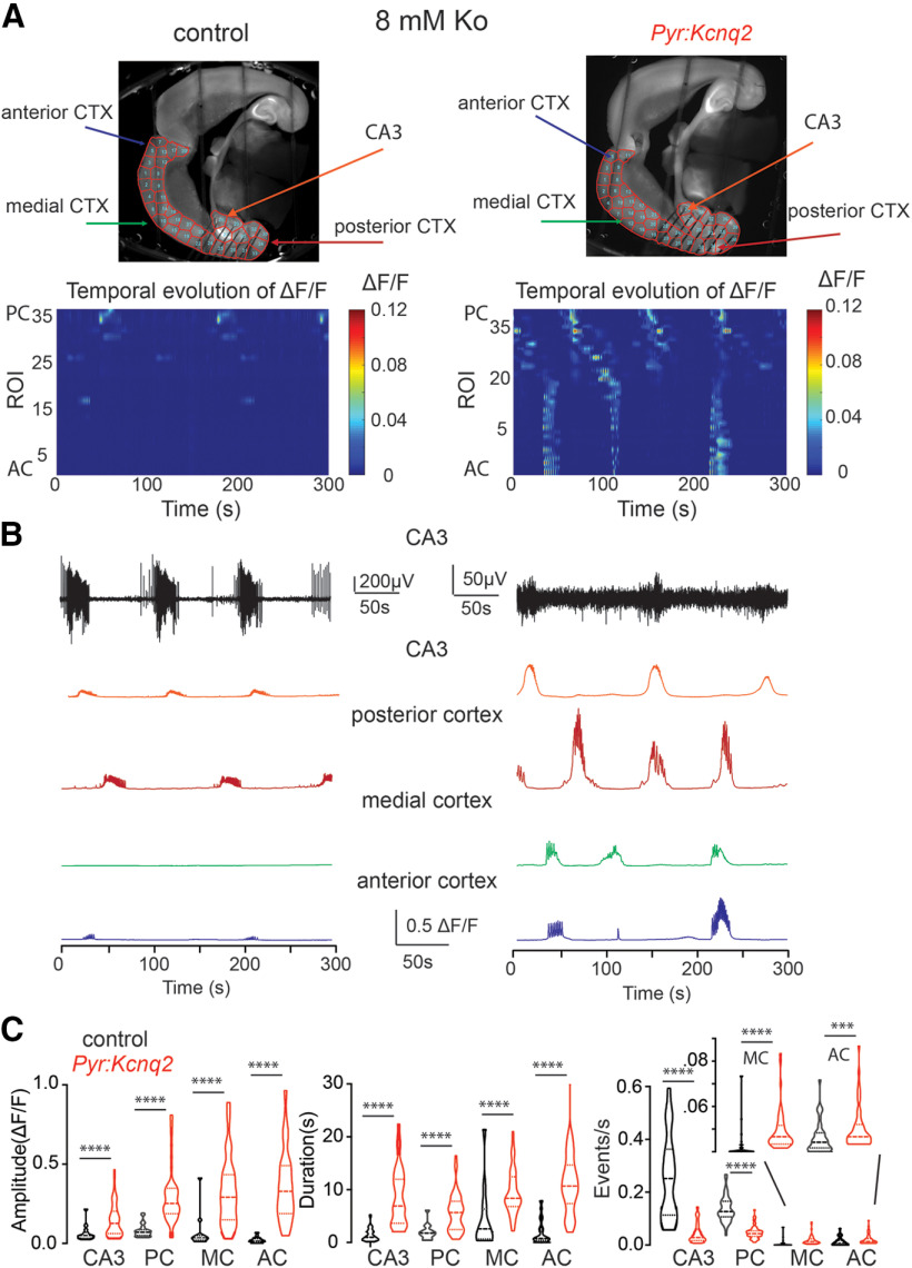
Deletion of Kcnq2 from excitatory neurons leads to elevated calcium activity across the forebrain in 8 mm Ko. A, top panels, Examples of acute slices from control and Pyr:Kcnq2 mice with one hemisphere segmented into ROIs. Bottom panels, 2D plots show the calcium activity across the different ROIs. The numbering corresponds to the segmented area shown on the top panels with lower values toward the AC and higher values toward the PC. Note that in the absence of Kcnq2, substantial calcium activity is measured across all regions of the forebrain. B, top two panels, Temporal evolution of the LFPs and ΔF/F recorded in parallel in the CA3 region of the hippocampus. Note that in contrast to the LFPs in slices from Pyr:Kcnq2 mice, the calcium responses are large and long lasting. Middle and bottom panels, Temporal evolution of the ΔF/F across multiple ROIs. C, Violin plots show the effect of Kcnq2 deletion on the amplitude, duration, and frequency of the calcium events for different anatomic regions. MC refers to the medial cortex. Note that ablation of Kcnq2 led to a large and uniform increase of the calcium response amplitude and duration. ****p < 0.0001. Additional details on the statistical analysis and number of replicates for this figure are found in Table 1 under the Figure 2 section.

