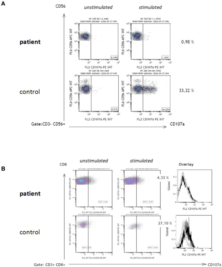
Figure 2.

CD107a degranulation assay. Degranulation of CD56+ NK-cells (A) and CD8+ CTL (B) was examined as previously published7. In brief, CD107a-expression on cell surfaces was analyzed by flow-cytometry in resting cells (A, B; left dot plot diagrams) and subsequent to 48 h stimulation with interleukin 2 (IL-2) of NK-cells (A, right dot plot diagrams), and phytohemagglutinin (PHA)/IL-2 of CD8+ T-cells (B, right dot plot diagrams), respectively.
