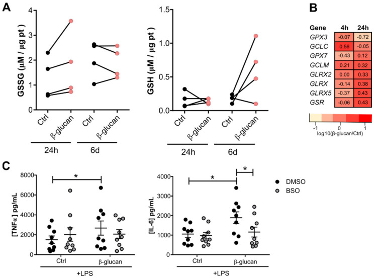
Figure 2.
Glutathione levels are modulated upon β-glucan exposure. (A) Reduced and oxidized glutathione intracellular levels in monocytes after 24 h exposure with 1 µg/mL β-glucan and 5 days after the resting period (n = 4 donors, pooled from two independent experiments). (B) Expression of genes involved in glutathione metabolism in monocytes exposed to β-glucan for 4 h and 24 h. Expression presented as log10(ratio). (C) TNFα and IL-6 produced by β-glucan-trained macrophages after a 1 h pretreatment with 100 µM BSO (n = 9 donors, pooled from three independent experiments, * p < 0.05 two-way ANOVA, Sidak’s multiple comparisons test) (mean ± SEM).

