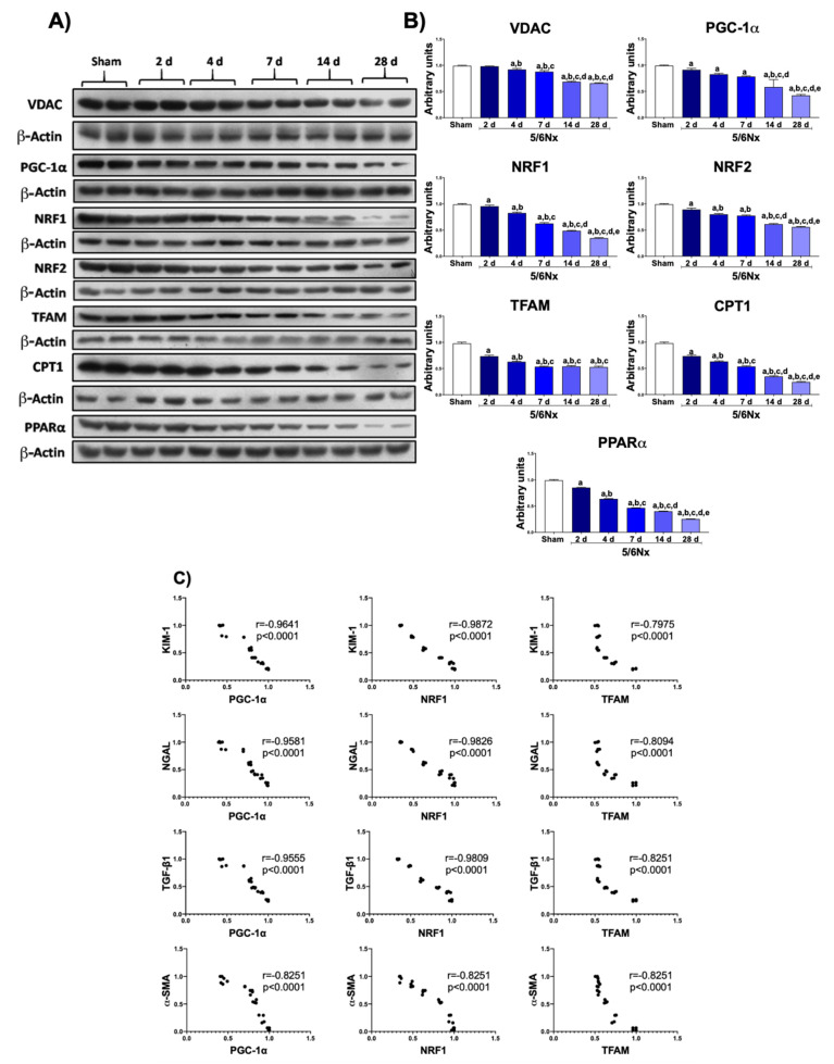
Figure 2.
Progressive decrease in mitochondrial biogenesis in remnant renal mass. (A) Western blots and (B) quantifications of mitochondrial proteins: voltage-dependent anion channel (VDAC), peroxisome proliferator-activated receptor gamma coactivator 1-alpha (PGC-1α), nuclear respiratory factor 1 (NRF1) and 2 (NRF2), mitochondrial transcription factor A (TFAM), carnitine palmitoyltransferase 1 (CPT1), and peroxisome proliferator-activated receptor alpha (PPARα). β-Actin was used as loading control. (C) Correlation analyses of kidney injury molecule-1 (KIM-1), neutrophil gelatinase-associated lipocalin (NGAL), profibrotic molecule transforming growth factor beta 1 (TGF-β1), and alpha smooth muscle actin (α-SMA) with PGC-1α, NRF1, or TFAM. Data are the mean ± SEM, n = 4. Tukey test. a = p ≤ 0.05 vs sham, b = p ≤ 0.05 vs. 2 days, c = p ≤ 0.05 vs. 4 days, d = p ≤ 0.05 vs. 7 days, e = p ≤ 0.05 vs. 14 days, 5/6Nx = 5/6 nephrectomy, d = days after 5/6Nx, sham = simulated operation/control group.

