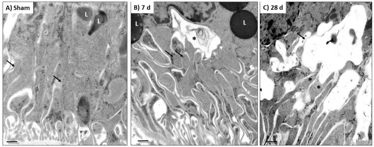
Figure 4.
Representative ultrastructural micrographs of epithelial cells from proximal convoluted tubules in (A) sham animals and in (B,C) rats with 5/6 nephrectomy (5/6Nx). (A) Normal appearance of the mild and basal cytoplasmic areas of an epithelial tubular cell from a sham animal, showing long mitochondria (arrows) and round electron dense lysosomes (L). (B) After 7 days (d) of 5/6Nx, there were small and round mitochondria (arrows) and large cytoplasmic vacuoles limited by a double membrane corresponding to autophagosomes (asterisk), which were near to mitochondria and large lysosomes (L). (C) After 28 days of kidney resection, these cytoplasmic double-membrane vacuoles (arrow) were larger and directly connected to mitochondria (asterisks). Scale magnification bar = 500 nm.

