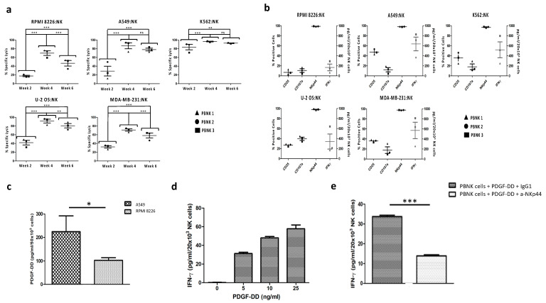
Figure 2.
Cytotoxicity of expanded PBNK cells and expression of cytotoxicity-associated molecules. (a) The cytotoxicity of three expanded PBNK cell donors towards RPMI 8226, A549, K562, U-2 OS, and MDA-MB-231 measured using the bioluminescence imaging assay at a 1:1 effector-to-target ratio for 24 h. (b) Expression of cytotoxicity-associated molecules (CD25, CD107a, NKp44, and IFN- γ) in PBNK cells in the fourth week of expansion after coculture with the different cancer cell lines (RPMI 8226, A549, K562, U-2 OS, and MDA-MB-231) at a 1:1 effector-to-target ratio for 24 h. (c) Expression of PDGF-DD in A549 and RPMI 8226. (d) Histogram with IFN-γ expression by PBNK cells after stimulation with different concentrations of PDGF-DD. (e) Graphic with IFN-γ expression by PBNK cells after stimulation with 5 ng/mL of PDGF-DD and IgG1, and after stimulation with 5 ng/mL of PDGF-DD and the anti-NKp44 mAb. Data in (a,b) are means ± SEM of the three expanded PBNK cell donors. Data in (c–e) are presented as means ± SD from three technical replicates of a PBNK cell pool. ns, not significant, *, p < 0.05, **, p < 0.01, ***, p < 0.001, one-way ANOVA, repeated measures test and Student’s t-test.

