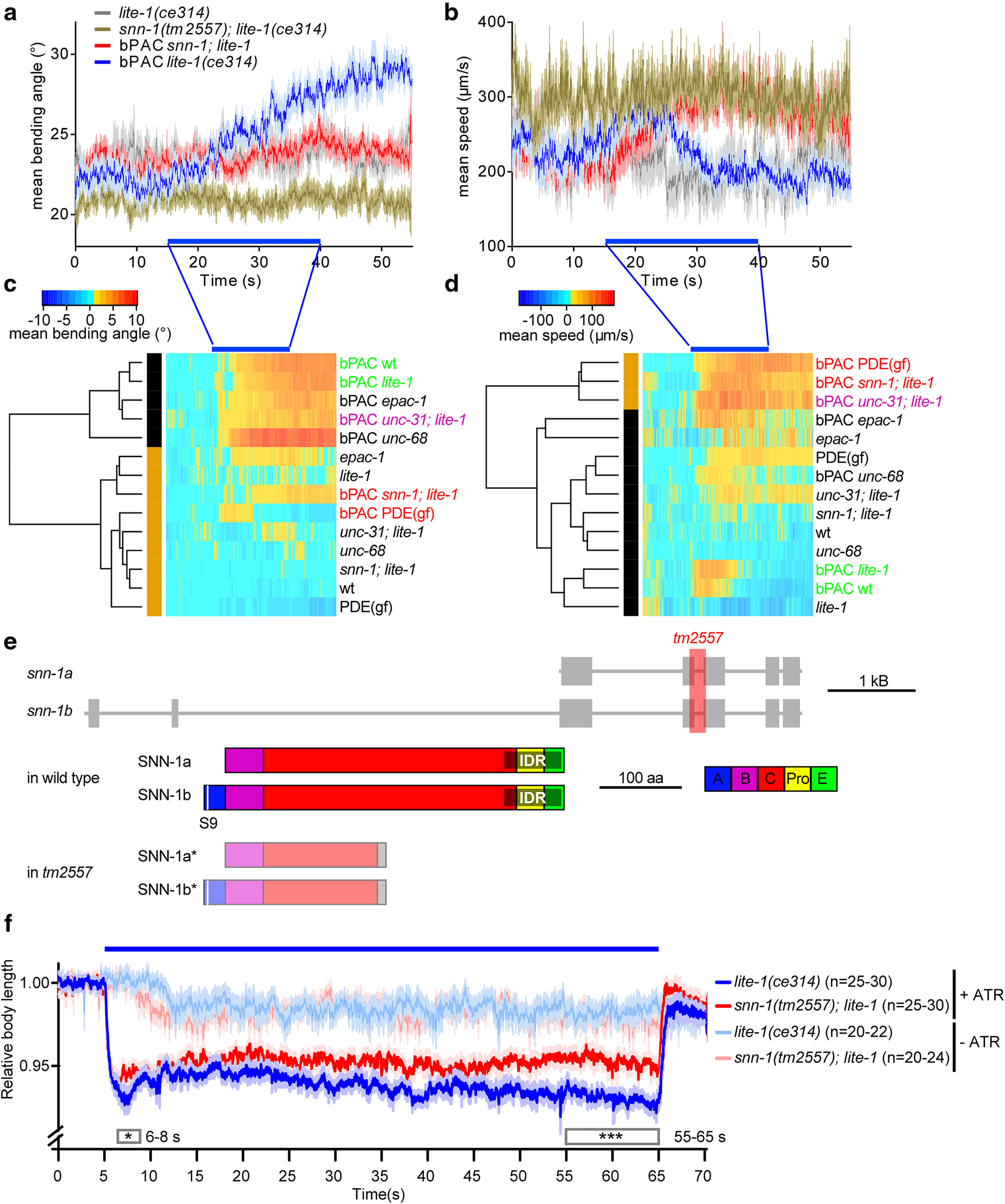
Figure 1.

Behavioral phenotypes induced by bPAC and ChR2 photostimulation in cholinergic neurons uncover synapsin as a target of cAMP increase and as a mediator of evoked release. bPAC was expressed in cholinergic neurons, and light effects on locomotion behavior were analyzed after video microscopy of individual animals. a, Mean (±SEM) bending angles (n ≥ 29), measured for 11 angles defined by 13 points along the body or (b) velocity of animals before, during, and after blue light stimulation (blue bar). Animals of the indicated genotypes were assessed. Bending angles (c) and velocities (d) for the indicated genotypes and transgenes were normalized to the mean before light stimulation and compared by dynamic cluster analysis. Two clusters are observed for both behaviors: one with wt animals (green), and another with a gain-of-function phosphodiesterase (PDE(gf)) and snn-1 mutants (red). e, C. elegans snn-1 gene on chromosome IV (boxes represent exons; lines represent introns), and protein structure of the a and b splice variants. Domains A, B, C, Pro (proline-rich), E, named by homology to the mammalian isoforms (Sudhof et al., 1989). Domains A and E are required for SV and synapsin oligomerization, whereas domain C interacts with the SV membrane. The intrinsic disordered region (gray shade) mediating phase separation of synapsin and SVs (Milovanovic et al., 2018) was annotated based on the Predictor of Natural Disordered Regions (www.pondr.com). S9, main PKA phosphorylation site. f, Body contraction in response to cholinergic photostimulation (ChR2). Mean (±SEM) normalized body length, number of animals, genotype as indicated. +/− ATR indicates incubation in all-trans retinal, the obligate ChR2 cofactor. Blue bar represents photostimulation. Boxes represent periods for which datasets were statistically significantly different. ***p < 0.001; *p < 0.05; two-way ANOVA and Bonferroni correction.
