Figure 5.
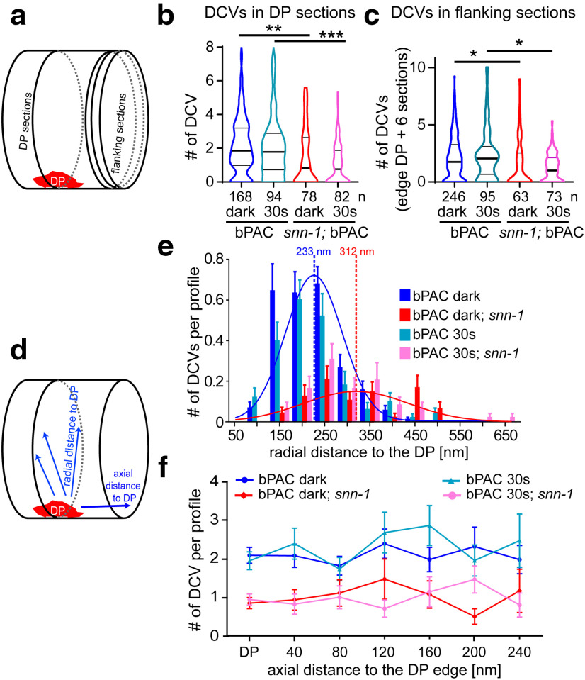
DCVs are largely depleted in snn-1(tm2557) synapses and distribute differently compared with wt. a, Sections analyzed by HPF-EM either contain the DP or are flanking the region containing the DP. b, DCVs per profile containing the DP plus two flanking sections in snn-1(tm2557) compared with wt, without and with 30 s photostimulation. Data shown as median and 25/75 quartiles (thick and thin lines), min to max. c, Same as in b, but in DP-flanking sections. d, Abundance of DCVs was analyzed either radially within a section, in distinct distances to the DP, or along the axon. e, Abundance of DCVs in distinct radial distances of DCVs relative to DP, quantified in 50 nm bins in untreated and 30 s stimulated wt and snn-1(tm2557) synapses. f, DCV abundance in sections containing the DP, or in sections of the indicated axial distance to the DP. b, c, e, f, Data are mean ± SEM. *p ≤ 0.05; **p ≤ 0.01; ***p ≤ 0.001; one-way ANOVA with Tukey correction.

