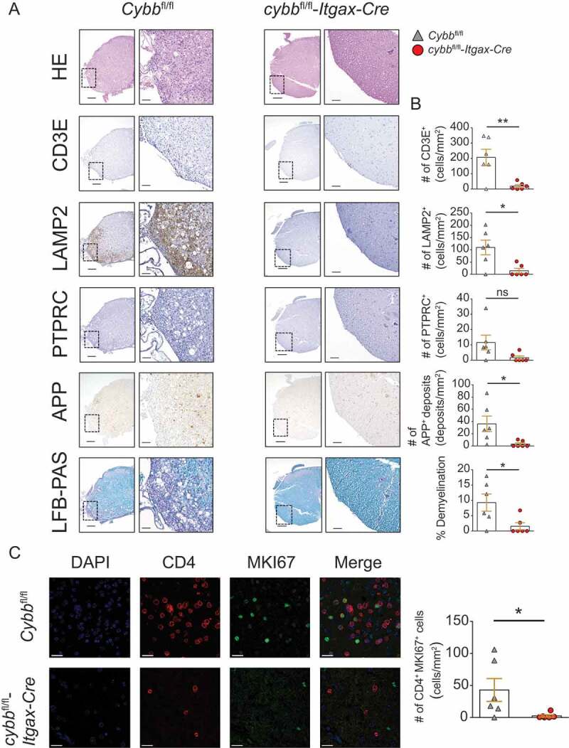
Figure 3.

Reduced immune cell infiltration, demyelination, and axonal damage in the CNS of cybbfl/fl-Itgax-Cre animals upon adoptive transfer EAE. (A) Histology of lumbar spinal cord sections from Cybbfl/fl and cybbfl/fl-Itgax-Cre animals on day 28 upon adoptive transfer of encephalitogenic CD4+ T cells using hematoxylin and eosin staining (HE), staining with antibodies against CD3E for T lymphocytes, against LAMP2 for myeloid cells, against PTPRC for B cells, against amyloid precursor protein for APP deposits and luxol fast blue (LFB-PAS) for demyelination. Scale bars: 200 µm (insert 50 µm) (left). (B) Quantification of cells or deposits per mm2 and fraction of demyelination is depicted (right). (C) Immunofluorescence microscopy analysis of CD4+ MKI67+ cells. Scale bars: 20 µm. Each data point represents one individual animal (Cybbfl/fl: gray triangles; cybbfl/fl-Itgax-Cre: red circles). Statistical analysis: Unpaired two-tailed Student t test was applied. Mean ± SEM is depicted. ns, not significant: P > 0.05; *P < 0.05; **P < 0.005
