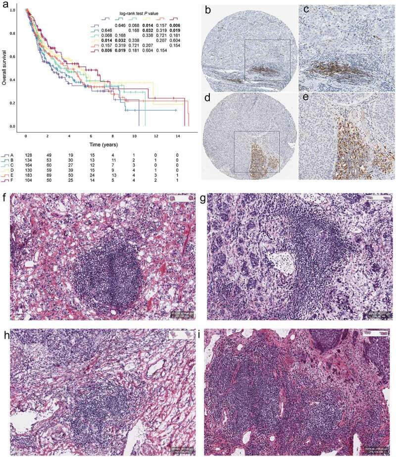
Figure 2.

Tertiary lymphoid structures in bladder cancers. (a) Kaplan–Meier curves for OS of 14 cohorts showing the association between MICs and OS. Representative images of tertiary lymphoid structures (TLSs) detected in formalin- fixed paraffin-embedded bladder cancers sections by immunohistochemistry staining showing CD20+ (brown) B cell zones (b and c) and LAMP+ (brown) DC cell zones (d-e). TLSs could be recognized as “small” lymph node like structures in HE slides of MIBC from TCGA (f-i)
