
Figure 3.
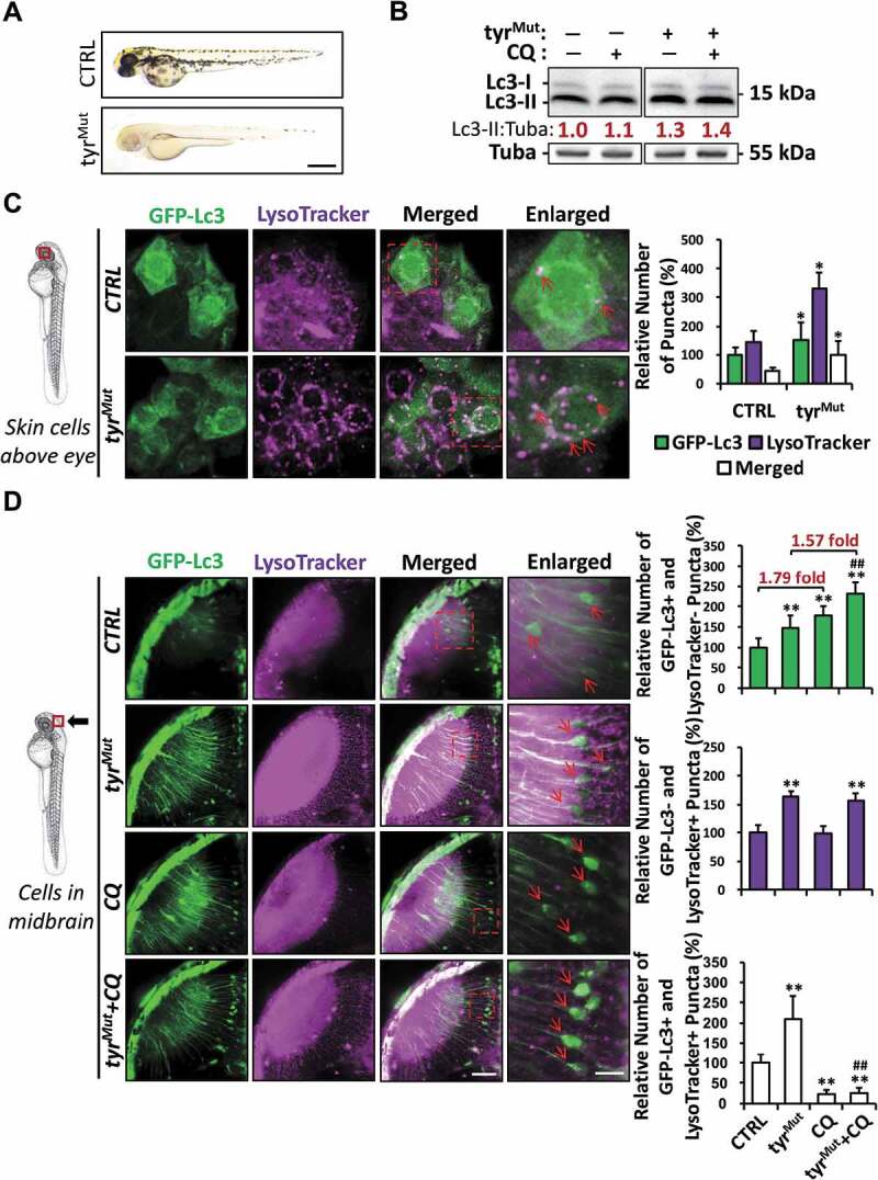
Aberrant autophagosome and autolysosome formation in tyrMut zebrafish embryos. (A) Representative bright-field images showing the pigmentation of 2 dpf zebrafish embryo injected with tyr gRNA+Cas9 protein (tyrMut) and their sibling control (CTRL). Scale bar: 0.5 mm. (B) Western blot results showing the level of Lc3-I and Lc3-II proteins in CTRL and tyrMut groups treated with chloroquine (CQ). Mean relative ratio of Lc3-II:Tuba was presented under the bands. 50 embryos were collected per group for three independent experiments. Two-way ANOVA with Tukey post hoc was applied, and a significant increase (p < 0.05) of Lc3-I and Lc3-II were detected in tyrMut compared with CTRL while no significant differences (p > 0.05) in Lc3-II:Tuba were detected between tyrMut treated with CQ and tyrMut. (C) Schematic diagram showing the position (periderm or basal epidermal cells above the eye) of imaging. Representative images of nine tyrMut Tg(GFP-Lc3) zebrafish embryos stained with LysoTracker from three independent experiments were shown. Three independent areas were selected from individual animals, and the relative number of GFP-Lc3+, LysoTracker+ and Merged (GFP-Lc3+ and LysoTracker+) puncta per cell were quantified. Red arrowhead, GFP-Lc3+ and LysoTracker+ puncta. *, p < 0.05 compared with CTRL. Scale bar: 10 μm (Merged), 5 μm (Enlarged). (D) Schematic diagram showing the position (cells in the midbrain) of imaging. The relative number of GFP-Lc3+ and LysoTracker−, GFP-Lc3− and LysoTracker+, and GFP-Lc3+ and LysoTracker+ puncta per cell in neurons of midbrain were counted based on Z-Stack (10 layers out of 100 layers) images. Representative images of nine tyrMut Tg(GFP-Lc3) zebrafish embryos treated CQ and stained with LysoTracker from three independent experiments were shown. Red arrowhead, GFP-Lc3+ and/or LysoTracker+ puncta. **, p < 0.01 compared with CTRL; ##, p < 0.01 compared with tyrMut. Scale bar: 40 μm (Merged), 3 μm (Enlarged)