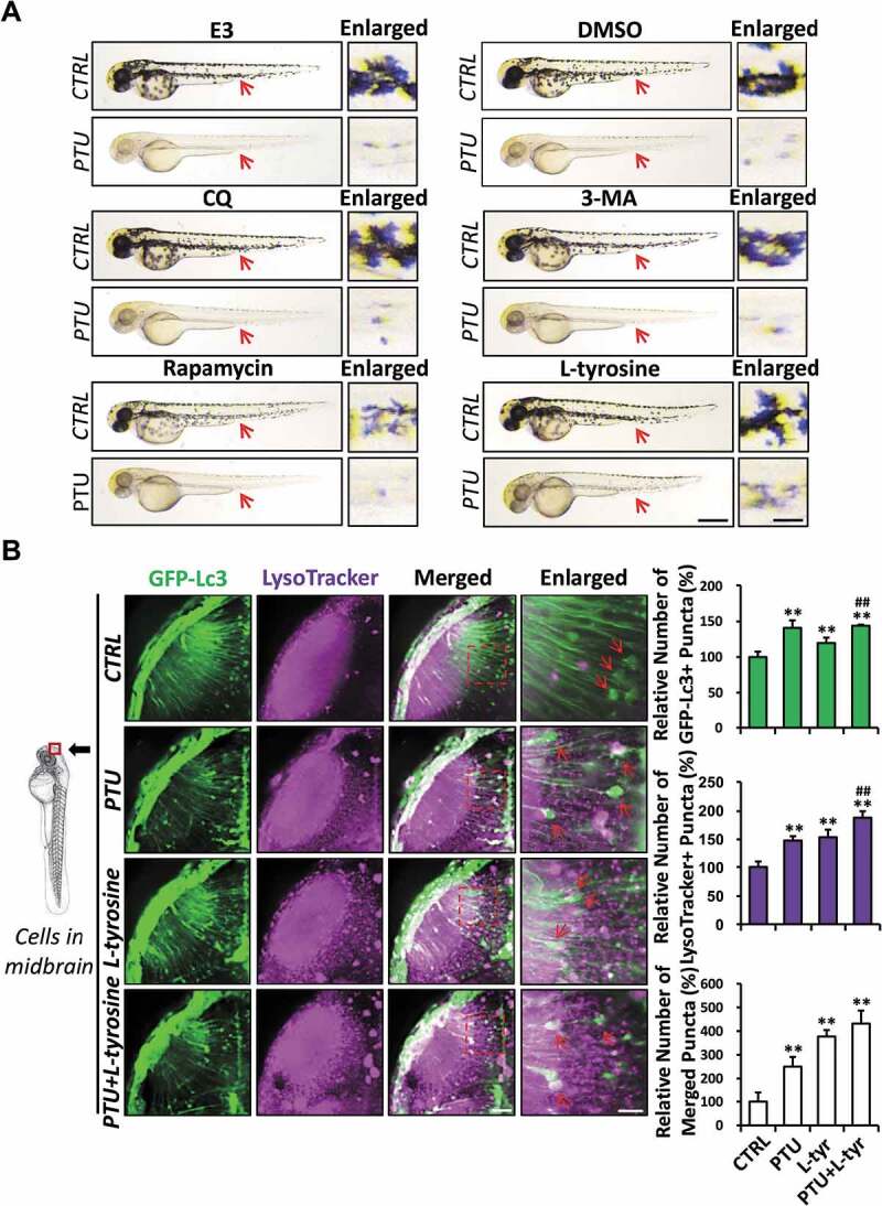
Figure 4.

L-tyrosine, but not autophagic modulators, restores melanogenesis in PTU-treated zebrafish embryos. (A) Representative bright-field images showing the pigmentation of 2 dpf zebrafish embryo treated with autophagic modulators, including E3, dimethyl sulfoxide (DMSO), chloroquine (CQ), 3-methyladenine (3-MA), rapamycin and L-tyrosine with or without PTU. Red arrowhead, melanin shown in Enlarged. Scale bar: 0.5 mm. (B) Schematic diagram showing the position (cells in the midbrain) of imaging. The relative number of GFP-Lc3+, LysoTracker+, and Merged (GFP-Lc3+ and LysoTracker+) puncta per cell were counted based on Z-Stack (10 layers out of 100 layers) images. Representative images of nine Tg(GFP-Lc3) zebrafish treated with PTU, L-tyrosine, or PTU+L-tyrosine and stained with LysoTracker prior to imaging from three independent experiments were shown. Red arrowhead, GFP-Lc3+ and/or LysoTracker+ puncta. **, p < 0.01 compared with CTRL; ##, p < 0.01 compared with L-tyrosine. Scale bar: 40 μm (Merged), 6 μm (Enlarged)
