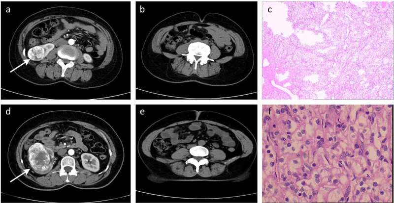
Figure 2.

Representative radiographic and histological images of patients. (a-c) Low-T-stage ccRCC in a 49-year-old woman. (d-f) High-T-stage ccRCC in a 54-year-old woman. (a) Enhanced CT showing right renal tumour (white arrow). (b) CT image with a relative visceral fat area (rVFA) of 18.2%. (c) Histologic photomicrograph confirming that the tumour did not involve capsule so as to be T1 stage. (d) Enhanced CT showing right renal tumour (white arrow). (e) CT image with a relative visceral fat area (rVFA) of 35.0%. (f) Histological micrographs confirming that the tumour involved the capsule, but was confined to the perirenal fascia so as to be T3 stage
