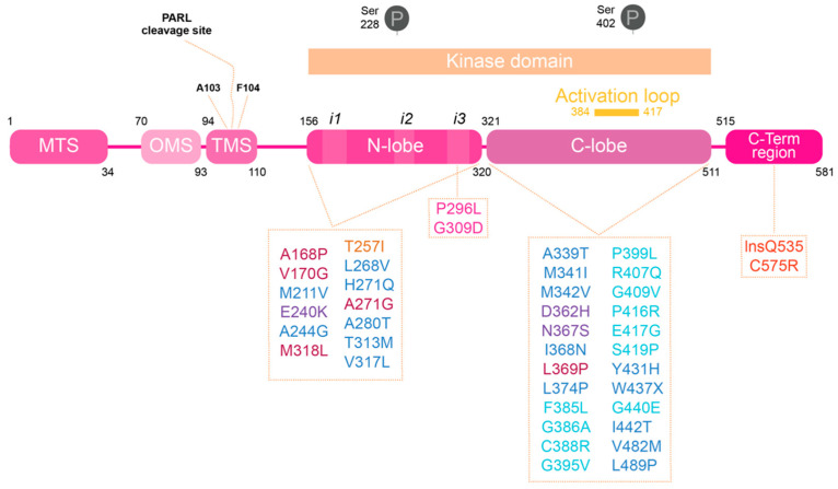
Figure 1.
Human PINK1 domains and PD associated mutations. PINK1 is divided into different regions. At the N-terminal are the regions responsible for the processing and delivery of PINK1 to mitochondria: mitochondrial targeting sequence (MTS), the region recently called named the mitochondria membrane localization signal (OMS) and the transmembrane sequence (TMS). Within the TMS resides the PARL cleavage sites. The kinase domain is divided into an N-lobe and C-lobe; it is also the PINK1 domain where the majority of PD-associated mutations are found, and where there are the well described phosphorylation sites [15]. At the N-lobe there are the different inserts (i1, i2 and i3) identified in bioinformatics studies performed using PINK1 insects structure. The activation loop, at the C-lobe, changes the proteins conformation from inactive to active state upon phosphorylation. PINK1-PD mutations can be divided into mutations that affect PINK1’s structure, kinase activity or substrate binding, depending on residues and protein regions affected: ATP binding pocket (bordeaux), kinase core (dark blue), catalytic mutations (purple), insert 2 (yellow), insert 3 (pink), activation loop (light blue) and C-terminal region (red).

