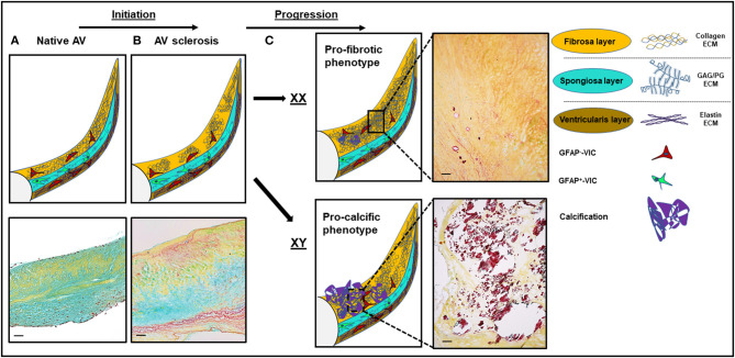
Figure 1.
Structural and histological changes in calcific aortic valve disease (CAVD). (A) The native aortic valve (AV) has three distinct extracellular matrix (ECM) layers: the fibrosa (yellow), the spongiosa (turquoise), and the ventricularis (dark yellow). VICs in the spongiosa layer express GFAP (48). (B) Disease initiation in CAVD is marked by fibrotic ECM expansion and disarray in the fibrosa layer. (C) Disease progression is sex-dependent: a more profibrotic phenotype in women and a more procalcific phenotype in men. Movat staining: yellow, collagen; turquoise, proteoglycans; dark purple, calcification. Scale bars indicate 100 μm. GFAP, glial fibrillary acidic protein. GAG, glycoaminoglycans; PG, proteoglycans; XX, female; XY, male.

