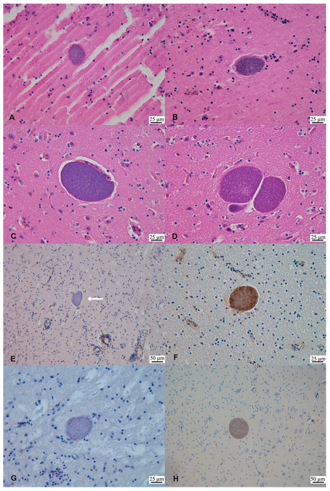
Figure 2.
Sarcocystis-like tissue cysts in the brain of striped dolphins (Stenella coeruleoalba) SD1 and SD2 from Liguria, Italy. (A) Parietal cortex (SD1). Protozoan tissue cyst measuring 70 × 50 µm. H&E. (B) Occipital cortex (SD1). Protozoan tissue cyst measuring 44.6 × 58.1 µm. H&E. (C) Frontal cortex (SD2). Protozoan tissue cyst measuring 72.83 × 116.34 µm. H&E. (D) Basal ganglia (SD2). Protozoan tissue cysts measuring (left-right reading) 110 × 119.3 µm, 40 × 19.8 µm and 50 × 99.1 µm. H&E. (E) Mesencephalon (SD1). Negative immunostaining of a protozoan tissue cyst (arrow). Monoclonal Ab anti-S. neurona. (F) Cerebellum (SD1). Positive labeling of Sarcocystis-like tissue cyst. Polyclonal Ab anti-S. falcatula. (G) Mesencephalon (SD1). Positive labeling of a protozoan tissue cyst bradyzoites. Polyclonal Ab anti-S. neurona. (H) Parietal cortex (SD2). Positive immunostaining of a protozoan tissue cyst bradyzoites. Polyclonal Ab anti-S. neurona.

