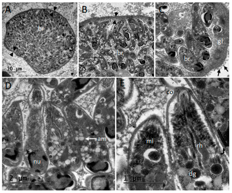
Figure 3.
Transmission electron microscopy micrographs of the Toxoplasma gondii tissue cyst studied from central nervous system of striped dolphin (Stenella coeruleoalba) SD2 in Liguria, Italy. (A) Section of the thin-walled tissue cyst; note the cyst wall (arrowheads) and densely packaged bradyzoites (br). (B,C) Details of the simple and thin cyst wall (arrowheads) and granular layer (gl) presenting vesicles (arrows). (D,E) Ultrastructural details of bradyzoites, note: nucleus (nu), micronemes (mi), dense granules (dg), amylopectin granules (am), conoid (co), and rhoptries (rh).

