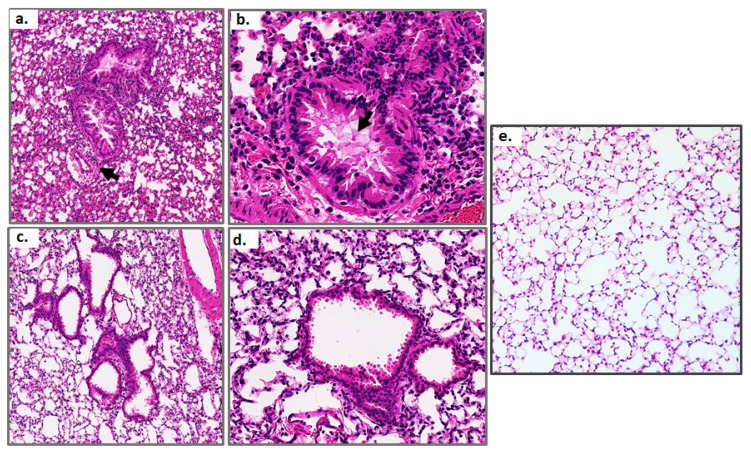
Figure 4.
Lung histology at day 7 and day 21 of a sublethal PVM infection compared to uninfected control. Hematoxylin and eosin-stained lung tissue sections from mice inoculated with a sublethal dose of PVM and evaluated on day 7 ((a,b); original magnifications, 20× and 64×, respectively) and day 21 ((c,d); original magnifications, 20× and 64×, respectively); control lung tissue (e) at original magnification of 20×. Arrow in (a) indicates neutrophils surrounding a blood vessel adjacent to an airway; arrow in (b) points to pulmonary edema within the large airway. Each image is representative of findings from n = 5 mice.

